文/李元昆 教授|新加坡國立大學楊潞齡醫學院,微生物與免疫學系/外科學系。
臨床及基礎研究中,已有越來越多證據顯示人類腸道微生物菌相 (gut microbiome) 能調節免疫系統、心智及其他生理功能的發育 (1-8)。成熟的人類腸道微生物菌相可分為兩大操作類型,分別是擬桿菌屬-雙歧桿菌屬型 (Bacteroides-Bifidobacterium type) 和普雷沃氏菌屬型 (Prevotella type) ,主要決定於飲食、地理位置以及生活型態(9-13)。擬桿菌屬-雙歧桿菌屬型主要分布於歐洲、北美洲和東亞 (9, 13–15),普雷沃氏菌屬型則是常見於東南亞、蒙古與非洲 (9, 14, 16)。母親會在懷孕與生產的過程中將自己的微生物菌相轉移給孩子 (17–20)。在嬰兒出生後,微生物菌相在母嬰之間的垂直轉移主要發生在腸道、產道、母乳、口腔以及皮膚等部位 (20–27)。西方國家自然產的新生兒,經由產道傳遞,會和母親有相同的腸道微生物菌相類型,也就是擬桿菌屬-雙歧桿菌屬型 (21, 26–29)。實際上不同菌種之間的相對豐富度在滿周歲前仍會改變 (23, 28, 30–32)。這些研究結果暗示,這類能「遺傳」到嬰兒的腸道微生物菌相組成,或許能夠保護兒童未來的健康。
母親只給了寶寶腸道微生物藍圖,並不決定其終其一生的發展
諷刺的是,這種常見於西方國家微生物菌相型的優勢菌:擬桿菌屬 (Bacteroides) 卻被列為許多已開發國家常見疾病的獨立風險因子,包含心血管疾病 (24, 25)、第二型糖尿病 (26, 27)、大腸直腸癌 (28-30)、心肌病變 (31)、類風溼性關節炎 (32)、發炎型腸道疾病 (33)、帕金森氏症 (34)、乳糜瀉 (35) 以及阿茲海默症 (36)。另外一方面,普雷沃氏菌屬(Prevotella) 為東南亞居民和素食者腸道主要菌群,也有直接或間接的證據暗示其與慢性發炎型疾病有關,包括牙周病、陰道菌叢變化症 (bacterial vaginosis) (37)、扁桃腺炎(38)、非酒精性脂肪肝引發的後期肝纖維化 (39)、心血管代謝疾病風險 (40) 以及氣喘 (41)等。 這些研究導向一個值得思考的重要健康問題:難道我們一生的健康與可能得到的疾病竟然被連結到出生那一刻?在回答這個問題之前,我們必須先了解決定腸道微生物菌相發展與建立的幾個關鍵因子。
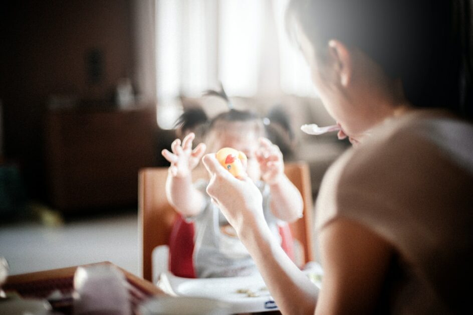
如上所述,自然產嬰兒,其第一時間接觸的微生物菌相來自於母親的垂直傳遞。因此,有人可能會假設我們的健康狀況或許在這個時刻就已經被母親「設定」了。令人意外地是,一份近期於東南亞的研究結果顯示,新生兒至離乳前,其腸道微生物菌相屬於擬桿菌屬-雙歧桿菌屬型,並非他們母親所擁有的普雷沃氏菌屬型 (42)。且這些嬰兒的腸菌相,會在離乳過程中逐步轉變成與母親相同的普雷沃氏菌屬型 (12, 14)。

剖腹產也會影響寶寶的腸胃道微生物菌相?
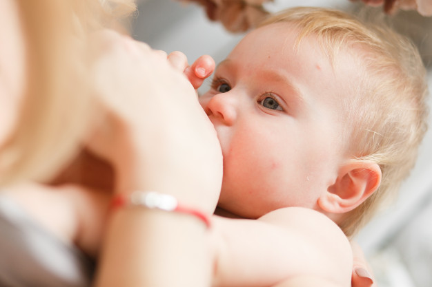
這項於印尼的研究,發現擬桿菌屬、雙岐桿菌屬 (Bifidobacterium) 與腸細菌科 (Enterobacteriaceae) 是嬰幼兒糞便中佔比最高的細菌,和過去於西方國家的研究結果相同 (23, 28–30)。在這群印尼嬰幼兒的糞便中,菌相隨著年紀的變化,主要來自於年紀較長的嬰幼兒糞便出現了一些特定的菌屬:在 6-12 月齡和 12-24 月齡的兩組中,主要是普雷沃氏菌屬、布勞特氏菌屬 (Blautia)、普拉梭菌屬 (Faecalibacterium)、毛螺菌科 (Lachnospiraceae) 以及瘤胃菌科 (Ruminococcaceae)等。同時,擬桿菌屬、雙岐桿菌屬、腸細菌科與克雷伯氏菌屬 (Klebsiella) 的相對豐富度則在離乳後逐漸下降 (6-12 月齡時),並在 24–48 月齡時持續維持較低的比率。在出生後短短不到一個月內,擬桿菌屬和雙岐桿菌屬就能以母親菌相的少數菌之姿,迅速在嬰兒的腸道內建立優勢微生物菌相,顯示新生兒的腸道環境可能非常適合這類菌屬的定殖與生長。同時卻又讓我們思考,為什麼剖腹產嬰兒即使是由母親哺乳,腸道內的擬桿菌屬和雙岐桿菌屬卻生長速度較為緩慢呢 (28, 43)?過去曾認為,這個差異可能是來自於剖腹產胎兒與母親有較少的垂直傳遞 (28, 43),然而根據這份印尼的研究,嬰兒早期腸道微生物菌相的快速建立,未必需要高量菌數之殖入。一個可能的解釋是,剖腹產手術過程會影響腸道的環境,進而阻撓共生菌在嬰兒腸道的建立過程。人類乳汁中的寡醣類能幫助雙岐桿菌屬在嬰兒腸道中成長 (44–46),且母乳富含脂質 (47),會促進膽酸分泌,進一步抑制普雷沃氏菌屬,並促進擬桿菌屬和雙岐桿菌屬的生長 (48, 49)。確實在該篇研究中,印尼嬰兒離乳前的排泄物 (糞水) 中含有較多的初級膽酸 (primary bile acids)。 離乳後的腸道微生物菌相改變也使得初級膽酸被轉換成次級膽酸 (secondary bile acids),與過去日本的研究結果相符 (42, 50)。離乳前嬰兒腸道中擬桿菌屬和雙岐桿菌屬的功能以及它們之所以佔據菌相多數的原因,值得注意。以該篇印尼研究來說,擬桿菌屬和雙岐桿菌屬不太可能與成人後的營養消化有關,因為在離乳後就會迅速轉成普雷沃氏菌屬主導的微生物相,且母親 (成人) 的腸道微生物菌相中擬桿菌屬和雙岐桿菌屬分布本就較低。在這篇研究中,與免疫細胞生長、成熟有關的細胞激素含量 (IL-1β, −2, −5, −8, −12, TNF-α 和 IFN-β) 也在離乳後上升。這也暗示了幼兒的主動免疫在離乳期開始發展。在一篇小鼠研究中, 腸道殖入Bacteroides fragilis (來自擬桿菌屬) 能夠抑制促發炎的TH17 免疫細胞反應 (51)。推測此篇印尼研究,離乳前擬桿菌屬的優勢定殖或許能抑制腸道中的促發炎細胞。在產後一個月內,普雷沃氏菌是母親陰道裡含量最豐富菌種。然而,即使生產過程中,嬰兒暴露於較多的普雷沃氏菌,但哺乳期間此菌的含量卻一直只在嬰兒的腸道微生物菌相佔少數。直到離乳後,嬰兒開始吃母乳以外的食物才逐漸轉為優勢菌種。有趣的是,歐洲與北美的母親,雖然腸道微生物菌相屬於擬桿菌屬-雙岐桿菌屬型,其陰道菌相卻仍以大量的普雷沃氏菌為主 (20, 23),即便歐美常見離乳後攝食的高脂/高蛋白飲食並不利於與此菌於腸道之繁衍 (12, 15)。儘管已開發國家飲食型態近數十年來有相當的演變,內源性因子所造就的普雷沃氏菌在母親陰道中的優勢,完全沒有受到影響。因為早期人類的飲食主要是以蔬菜為主,與現今的印尼族群飲食型態近似。然而,來自母體的微生物傳遞依舊被認為是嬰幼兒腸道微生物菌相的重要來源之一,因為所有孩子腸道中的菌種都可以在母親的微生物相中找到。不過,母親的微生物菌相中仍有許多額外的菌種,這表示只有少部分菌種可以透過腸道、陰道以及母乳傳遞給嬰兒,並且成功建立起嬰兒的微生物菌相。更重要的是,在母體優勢的菌種,並不一定在嬰兒腸道環境中也具相同的競爭優勢。這現在的其中一個解釋為,嬰兒的腸道環境可能偏好具有帶著某些特定功能基因的菌種 (27), 也有可能是除了垂直傳遞以外,菌相的水平傳遞也占了一席之地 (52)。無論如何,該篇研究最後也顯示嬰幼兒的腸菌相,會隨著成長而逐漸與母親的腸菌相愈來愈相近 (26, 28)。
這篇研究,使用了一個相關係數指標來評估腸道共生菌與一些已知致病菌之間的相關程度;此指標係將共生菌與潛在致病菌的相關係數予以加總,指標 0 表示任何共生菌與病原菌均不相關。這個指標在這篇印尼嬰兒的研究中,離乳前為 4.9,而在離乳後則是上升到 31.0。前後的數字變化顯示,離乳前的共生菌群和母乳中可提供被動免疫防護的抗體、免疫球蛋白能夠壓制並避免潛在致病菌的生長。這些共生菌會提供嬰兒保護力,直到離乳前後他們自己的免疫系統完整發育。這種微生物菌相也許是一個演化上的保護策略,能在較不注重衛生習慣的過去,或者是某些開發中國家的現今,提供較早離乳的嬰兒足夠的保護力。 霍亂弧菌為開發中國家最廣泛的致命腸道病原菌。

雙岐桿菌屬在各年齡層均與複雜碳水化合物、當地水果以及乳製品的攝取呈負相關。此印尼的研究結果也支持了先前的研究發現,飲食中抗性澱粉的存在會與雙岐桿菌屬數量呈現負相關(14)。印尼人的主食為秈米 (Indica rice),含有較多的抗性澱粉。印尼嬰兒離乳後攝取秈米,抗性澱粉持續存在於腸道中,造成腸道中游離膽酸被移除,進而使得普雷沃氏菌屬增殖,而降低雙岐桿菌屬 (10)。此外,普雷沃氏菌屬能夠發酵碳水化合物 (14, 15),攝食高碳水化合物的印尼人腸道環境有利其增殖。再者,研究也發現當地的水果及乳製品似乎會抑制雙岐桿菌屬的生長,且具有強烈的劑量效應。當地的果樹為了在高溫、容易滋生細菌的環境下生長,會產生抗菌的化學物質 (54–56)。而雙岐桿菌屬是否屬於對這類抗菌化學物質敏感的微生物,還需要進一步的研究證實。若屬實,這可能就是印尼幼兒離乳後腸道雙岐桿菌屬急遽下降的原因,同時也可解釋為何母親的腸道微生物菌相含有較少雙岐桿菌屬。在某些幼兒攝取較為西化飲食 (速食) 的案例中,來自麵包或洋芋片的可消化碳水化合物,可能造成速食攝取與雙岐桿菌屬之間出現正相關 (14)。過去在歐洲、北美洲以及東亞兒童所觀察到的雙岐桿菌屬保健功能 (33-36, 43, 53),在印尼兒童或許是由別種共生菌所取代,例如腸球菌 (Enterococcus faecalis) (57, 58)。嬰幼兒成長到 24-48 月齡時,可能已發展出一套成熟平衡的腸道微生物菌相系統。或許因與母親的飲食日趨相似,其與母親的微生物菌相亦就非常雷同。
綜此,至今研究指出:
- 在出生僅僅一個月內,嬰兒體內優勢菌相即可相對快速的建立,其起始於來自母親極少菌量之殖入,但決定於嬰兒腸道環境因子,非母親菌相中含量最多的菌。
- 嬰兒離乳前的腸道優勢菌相與內外緣因子有關,包括膽酸濃度、細胞激素、母乳中寡醣以及黏液素的醣基 (mucin glycans) 等,這些因子有利於擬桿菌屬及雙岐桿菌屬成為優勢菌種。
- 在嬰兒離乳前,擬桿菌屬及雙岐桿菌屬的量與潛在致病菌的量呈負相關。唯其於抵抗傳染性疾病中所扮演的角色還需要更多臨床研究證實。
- 離乳後的飲食型態,驅動嬰兒糞便微生物菌相組成的改變,故離乳期代表了微生物菌相變化的過渡期窗口。
- 綜而言之,決定兒童腸道微生物菌相主要應是腸道環境和飲食組成,而非完全轉殖自母體。也就是說,離乳期的飲食為改善腸道微生物菌相的一個務實捷徑。
參考文獻
- Turroni F, Milani C, Duranti S, Lugli GA, Bernasconi S, Margolles A, Di Pierro F, van Sinderen D, Ventura M. The infant gut microbiome as a microbial organ influencing host well-being. Italian J Pediatrics. 2020;46:16.
- Olsza k T, An D, Zeissig S, Vera MP, Richter J, Franke A,Glickman JN, Siebert R, Baron RM, Kasper DL, et al. Microbial exposure during early life has persistent effects on natural killer T cell function. Science. 2012;336(6080):489–493. doi:10.1126/science.1219328.
- Gensollen T, Iyer SS, Kasper DL, Blumberg RS. How colonisation by microbiota in early life shapes the immune system. Science. 2016;352(6825):539–544. doi:10.1126/science.aad9378.
- Scheepers L, Penders J, Mbakwa CA, Thijs C, Mommers M, Arts I. The intestinal microbiota composition and weight development in children: the KOALA birth cohort study. Int J Obesity. 2015;39:16–25. doi:10.1038/ijo.2014.178.
- Milani C, Duranti S, Bottacini F, Casey E, Turroni F, Mahony J, Belzer C, Delgado Palacio S, Arboleya Montes S, Mancabelli L, et al. The first microbial colonizers of the human gut: composition, activities and health implications of the infant gut microbiota. Microbiol Mol Biol Rev. 2017;81(4):e00036–17.
- Mulligan CM, Friedman JE. Maternal modifiers of the infant gut microbiota: metabolic consequences. J Endocrinol. 2017;235(1):R1–R12. doi:10.1530/JOE-17-0303.
- Fujimura KE, Sitarik AR, Havstad S, Lin DL, Levan S, Fadrosh D, Panzer AR, LaMere B, Rackaityte E, Lukacs NW, et al. Neonatal gut microbiota associates with childhood multisensitised atopy and T cell differentiation. Nat Med. 2016;22(10):1187–1191. doi:10.1038/nm.4176.
- Ismail IH, Boyle RJ, Licciardi PV, Oppedisano F, Lahtinen S, Robins-Browne RM, Tang MLK. Early gut colonization by Bifidobacterium breve and B. catenulatum differentially modulates eczema risk in children at high risk of developing allergic disease. Paediatric Allergy Immunol. 2016;27(8):838–846. doi:10.1111/pai.12646.
- De Filippo C, Cavalieri D, Di Paola M, Ramazzotti M, Poullet JB, Massart S, Collini S, Pieraccini G, Lionetti P. Impact of diet in shaping gut microbiota revealed by acomparative study in children from Europe and rural Africa. Proc Natl Acad Sci USA. 2010;107(33):14691–14696. doi:10.1073/pnas.1005963107.
- Khine WWT, Zhang YW, Goie JY, Wong MS, Liong MT, Lee YY, Cao H, Lee Y-K. Gut microbiome of preadolescent children of two ethnicities residing in three distant cities. Sci Rep. 2019;9(1):78931. doi:10.1038/s41598-019-44369-y.
- Kisuse J, La-Ongkham O, Nakphaichit M, Therdtatha P, Momoda R, Tanaka M, Fukuda S, Popluechai S, Kespechara K, Sonomoto K, et al. Urban diets linked to gut microbiome and metabolome alterations in children: a comparative cross-sectional study in Thailand. Front Microbiol. 2018;9:1345. doi:10.3389/fmicb.2018.01345.
- Nakayama J, Yamamoto A, Palermo-Conde LA,Higashi K, Sonomoto K, Tan J, Lee Y-K. Impact of Westernized diet on gut microbiota in children on Leyte islands. Front Microbiol. 2017;8:197. doi:10.3389/fmicb.2017.00197.
- Yatsunenko T, Rey FE, Manary MJ, Trehan I, Dominguez-Bello MG, Contreras M, Magris M, Hidalgo G, Baldassano RN, Anokhin AP, et al. Human gut microbiome viewed across age and geography. Nature. 2012;486(7402):222–227. doi:10.1038/nature11053.
- Nakayama J, Yamamoto A, Palermo-Conde LA, Higashi K, Sonomoto K, Tan J, Lee Y-K. Diversity in gut bacterial community of school-age children in Asia. Sci Rep. 2015;5:8397. doi:10.1038/srep08397.
- Wu GD, Chen J, Hoffmann C, Bittinger K, Chen -Y-Y, Keilbaugh SA, Bewtra M, Knights D, Walters WA, Knight R, et al. Linking long-term dietary patterns with gut microbial enterotypes. Science. 2011;334:6052. doi:10.1126/science.1208344.
- Zhang JC, Guo Z, Lim AA, Zheng Y, Koh EY, Ho D, Qiao J, Huo D, Hou Q, Huang W, et al. Mongolians core gut microbiota and its correlation with seasonal dietary changes. Sci Rep. 2014;4:5001. doi:10.1038/srep05001.
- Pelzer E, Gomez-Arango LF, Barrett HL, Nitert MD. Review: maternal health and the placental microbiome. Placenta. 2017;54:30–37. doi:10.1016/j.placenta.2016.12.003.
- Rautava S, Luoto R, Salminen S, Isolauri E. Perinatal microbial contact and the origins of human disease. Nat Rev Gastroenterol Hepatol. 2012;9:565–576. doi:10.1038/nrgastro.2012.144.
- Penders J, Thijs C, Vink C, Stelma CV, Snijders B, Kummeling I, van den Brandt PA, Stobberingh EE. Factors influencing the composition of the intestinal microbiota in early infancy. Paediatrics. 2006;118(2):511–521. doi:10.1542/peds.2005-2824.
- Dominguez-Bello MG, Costello EK, Contreras M, Magris M, Hidalgo G, Fierer N, Knight R. W. W. T. KHINE ET AL. Delivery mode shapes the acquisition and structure of the initial microbiota across multiple body habitats in newborns. Proc Natl Acad Sci USA. 2010;107(26):11971–11975. doi:10.1073/pnas.1002601107.
- Asnicar F, Manara S, Zolfo M, Truong DT, Scholz M, Armanini F, Ferretti P, Gorfer V, Pedrotti A, Tett A, et al. Studying vertical microbiome transmission from mothers to infants by strain-level metagenomics profiling. mSystem. 2017;2(1):e00164–16. doi:10.1128/mSystems.00164-16.
- Funkhouser LJ, Bordenstein SR. Mom knows best: the universality of maternal microbial transmission. PLoS Biol. 2013;11(8):e1001631. doi:10.1371/journal.pbio.1001631.
- Mueller NT, Bakacs E, Combellick J, Grigoryan Z, Dominguez-Bello MG. The infant microbiome development: mom matters. Trends Mol Med. 2015;21(2):109–117. doi:10.1016/j.molmed.2014.12.002.
- Roger LC, Costabile A, Holland DT, Hoyles L, McCartney AL. Examination of fecal Bifidobacterium populations in breast- and formula-fed infants during the first 18 months of life. Microbiology Society. 2010;156(11):3329–3341. doi:10.1099/mic.0.043224-0.
- Koenig JE, Spor A, Scalfone N, Fricker AD, Stombaugh J, Knight R, Angenent LT, Ley RE. Succession of microbial consortia in the developing infant gut microbiome. Proc Natl Acad Sci USA. 2011;108(Suppl.1):4578–4585. doi:10.1073/pnas.1000081107.
- Ferretti P, Pasolli E, Tett A, Asnicar F, Gorfer V, Fedi S, Armanini F, Truong DT, Manara S, Zolfo M, et al. Mother-to-infant microbial transmission from different body sites shapes the developing infant gut microbiome. Cell Host Microbe. 2018;24:133–145. doi:10.1016/j.chom.2018.06.005.
- Yassour M, Jason E, Hogstrom LJ, Arthur TD, Tripathi S, Siljander H, Selvenius J, Oikarinen S, Hyöty H, Virtanen SM, et al. Strain-level analysis of mother-tochild bacterial transmission during the first few months of life. Cell Host Microbe. 2018;24:146–154. doi:10.1016/j.chom.2018.06.007.
- Bäckhed F, Roswall J, Peng Y, Feng Q, Jia H, KovatchevaDatchary P, Li Y, Xia Y, Xie H, Zhong H, et al. Dynamics and stabilization of the human gut microbiome during the first year of life. Cell Host Microbe. 2015;17:690–730. doi:10.1016/j.chom.2015.04.004.
- Makino H. Bifidobacterial strains in the intestines of newborns originate from their mothers. Biosci Microbiota Food Health. 2018;37:79–85. doi:10.12938/bmfh.18-011.
- Vaishampayan PA, Kuehl JV, Froula JL, Morgan JL, Ochman H, Francino MP. Comparative metagenomics and population dynamics of the gut microbiota inmother an infant. Genome Biol Evol. 2010;2:53–66. doi:10.1093/gbe/evp057.
- Milani C, Turroni F, Duranti S, Lugli GA, Mancabelli L, Ferrario C, van Sinderen D, Ventura M. Genomics of the genus Bifidobacterium reveals species-specific adaptation to the glycan-rich gut environment. Appl Environ Microbiol. 2015;82(4):980–991. doi:10.1128/AEM.03500-15.
- Duranti S, Lugli GA, Mancabelli L, Armanini F, Turroni F, James K, Ferretti P, Gorfer V, Ferrario C, Milani C, et al. Maternal inheritance of bifidobacterial communities and bifidophages in infants through vertical transmission. Microbiome. 2017;5:66. doi:10.1186/s40168-017-0282-6.
- Milani C, Duranti S, Bottacini F, Casey E, Turroni F, Mahony J, Belzer C, Delgado Palacio S, Arboleya Montes S, Mancabelli L, et al. The first microbial colonizers of the human gut: composition, activities and health implications of the infant gut microbiota. Microbiol Mol Biol Rev. 2017;81(4):e00036–17.
- 34. Mulligan CM, Friedman JE. Maternal modifiers of the infant gut microbiota: metabolic consequences. J Endocrinol. 2017;235(1):R1–R12. doi:10.1530/JOE-17-0303.
- Fujimura KE, Sitarik AR, Havstad S, Lin DL, Levan S, Fadrosh D, Panzer AR, LaMere B, Rackaityte E, Lukacs NW, et al. Neonatal gut microbiota associates with childhood multisensitised atopy and T cell differentiation. Nat Med. 2016;22(10):1187–1191. doi:10.1038/nm.4176.
- Ismail IH, Boyle RJ, Licciardi PV, Oppedisano F, Lahtinen S, Robins-Browne RM, Tang MLK. Early gut colonization by Bifidobacterium breve and B. catenulatum differentially modulates eczema risk in children at high risk of developing allergic disease. Paediatric Allergy Immunol. 2016;27(8):838–846. doi:10.1111/pai.12646.
- Wampach L, Heintz-Buschart A, Fritz JV, RamiroGarcia J, Habier J, Herold M, Narayanasamy S, Kaysen A, Hogan AH, Bindl L, et al. Birth mode is associated with earliest strain-conferred gut microbiome functions and immunostimulatory potential. Nat Commun. 2018;9:5091. doi:10.1038/s41467-018-07631-x.
- Fouhy F, Watkins C, Hill CJ, ÓShea C-A, Nagle B, Dempsey EM, ÓToole PW, Ross RP, Ryan CA, Stanton C. Perinatal factors affect the gut microbiota up to four years after birth. Nat Commun. 2019;10:1517. doi:10.1038/s41467-019-09252-4.
- Shao Y, Forster SC, Tsaliki E, Vervier K, Strang A, Simpson N, Kumar N, Stares MD, Rodger A, Brocklehurst P, et al. Stunted microbiota and opportunistic pathogen colonisation in caesarean-section birth. Nature. 2019;574:117–121. doi:10.1038/s41586-019-1560-1.
- Nogacka A, Salazar N, Suárez M, Milani C, Arboleya S, Solís G, Fernández N, Alaez L, Hernández-Barranco AM, de Los Reyes-gavilán CG, et al. Impact of intrapartum antimicrobial prophylaxis upon the intestinal microbiota and the prevalence of antibiotic resistance genes in vaginally delivered full-term neonates. Microbiome. 2017;5(1):93. doi:10.1186/s40168-017-0313-3.
- Neuman H, Forsythe P, Uzan A, Avni O, Koren O. Antibiotics in early life: dysbiosis and the damage done. FEMS Microbiol Rev. 2018;42:489–499. doi:10.1093/femsre/fuy018.
- Khine WWT, Rahayu ES, See TY, Kuah S, Salminen S, Nakayama J, Lee YK. Indonesian children fecal microbiome from birth until weaning was different from microbiomes of their mothers, Gut Microbes, doi: 10.1080/19490976.2020.1761240.
- Dzidic M, Boix-Amorós A, Selma-Royo M, Mira A, Collado MC. Gut microbiota and mucosal immunity in the neonate. Med Sci (Basel). 2018;6:E56.
- Aakko J, Kumar H, Rautava S, Wise A, Autran C, Bode L, Isolauri E, Salminen S. Human milk oligosaccharide categories define the microbiota composition in human colostrum. Benef Microbes. 2017;8(4):563–567. doi:10.3920/BM2016.0185.
- Marcobal A, Barboza M, Froehlich JW, Block DE, German JB, Lebrilla CB, Mills DA. Consumption of human milk oligosaccharides by gut-related microbes. J Agric Food Chem. 2010;58(9):5334–5340. doi:10.1021/jf9044205.
- Duranti S, Lugli GA, Milani C, James K, Mancabelli L, Turroni F, Alessandri G, Mangifesta M, Mancino W, Ossiprandi MC, et al. Bifidobacterium bifidum and the infant gut microbiota: an intriguing case of microbehost co-evolution. Environ Microbiol. 2019;21 (10):3683–3695. doi:10.1111/1462-2920.14705.
- Andreas NJ, Kampmann B, Mehring L-DK. Human breast milk: a review on its composition and bioactivity. Early Hum Dev. 2015;91(11):629–635. doi:10.1016/j. earlhumdev.2015.08.013.
- Hayashi H, Shibata K, Sakamoto M, Tomita S, Benno Y. Prevotella copri sp. Nov. and Prevotella stercorea sp. Nov., isolated from human feces. Int J Syst Evol Microbiol. 2007;57:941–946. doi:10.1099/ijs.0.64778-0.
- Wahlstrom A, Sayin SI, Marschall H-U, Backhed F. Intestinal crosstalk between bile acids and microbiota and its impact on host metabolism. Cell Metab. 2016;24(1):41–50. doi:10.1016/j.cmet.2016.05.005.
- Masaru. T, Sanefuji M, Morokuma S, Yoden M, Momoda R, Sonomoto K, Ogawa M, Kato K, Nakayama J. The association between gut microbiota development and maturation of intestinal bile acid metabolism in the first three years of healthy Japanese infants. Gut Microbes. 2019. doi:10.1080/19490976.2019.1650997.
- Round JL, Lee SM, Li J, Tran G, Jabri B, Chatila TA, Mazmanian SK. The Toll-like receptor 2 pathway establishes colonisation by a commensal of the human microbiota. Science. 2011;332:974–977. doi:10.1126/science.1206095.
- Moeller AH, Suzuki TA, Phifer-Rixey M, Nachman MW. Transmission modes of the mammalian gut microbiota. Science. 2018;362:453–457. doi:10.1126/science.aat7164.
- Kim Y-G, Sakamoto K, Seo SU, Pickard JM, Gillilland MG 3rd, Pudlo NA, Hoostal M, Li X, Wang TD, Feehley T, et al. Neonatal acquisition of Clostridia species protects against colonization by bacterial pathogens. Science. 2017;356:315–319. doi:10.1126/science.aag2029.
- Oyedemi BOM, Kotsia EM, Stapleton PD, Gibbons S. Capsaicin and gingerol analogues inhibit the growth of efflux-multidrug resistant bacteria and R-plasmids conjugal transfer. J Ethnopharmacol. 2019;245:111871. doi:10.1016/j.jep.2019.111871.
- Kariu T, Nakao R, Ikeda T, Nakashima K, Potempa J, Imamura T. Inhibition of gingipains and Porphyromonas gingivalis growth and biofilm formation by prenyl flavonoids. J Periodontal Res. 2017;52(1):89–96. doi:10.1111/jre.12372.
- Cushnie TP, Cushnie B, Lamb AJ. Alkaloids: an overview of their antibacterial, antibiotic-enhancing and antivirulence activities. Int J Antimicrob Agents. 2014;44:377–386. doi:10.1016/j.ijantimicag.2014.06.001.
- Are A, Aronsson L, Wang S, Greicius G, Lee Y-K, Gustafsson JA, Pettersson S, Arulampalam V. Enterococcus faecalis from newborn babies regulate endogenous PPARgamma activity and IL-10 levels in colonic epithelial cells. Proc Natl Acad Sci USA. 2008;105:1943–1948. doi:10.1073/pnas.0711734105.
- Wang SG, Hibberd ML, Pettersson S, Lee Y-K. Enterococcus faecalis from healthy infants modulates inflammation through MAPK signaling pathways. PLoS One. 2014;9(5):e97523. doi:10.1371/journal.pone.0097523.


